Nub Theory: Can the 12-Week Ultrasound Really Show Gender?
Yes, within real limits. Nub theory reads the angle of the genital tubercle (the "nub") on a 12-week ultrasound profile. Peer-reviewed studies show trained readers get it right roughly 87–92% of the time at 12 weeks, rising toward 98–100% by week 13. Before 11 weeks it's guesswork, and it only works when the scan captures the right view.
Unlike most gender folklore, this one has published research behind it, which is reason enough to look at the numbers.
What Is the Nub, Exactly?
Every baby, boy or girl, develops the same small structure between the legs during the first trimester: the genital tubercle. Until about week 11 it looks identical in everyone. It will later become either a penis or a clitoris, and at this stage what separates the two is the angle rather than the shape.
As development progresses, the tubercle in boys begins rotating upward, away from the spine, while in girls it stays roughly level. That divergence is the entire basis of nub theory: on a side-profile ultrasound around 12–13 weeks, the direction the nub points is a real anatomical signal, unlike heart rate or bump shape.
How to Read the Nub Angle
You need a mid-sagittal profile, meaning the baby lying sideways with the spine visible, the classic "NT scan" pose from the 12-week appointment. Then:
- Find the line of the lower spine (the lumbosacral skin surface).
- Find the nub, a small protrusion between the legs, at the baby's rump end.
- Compare the nub's direction with the spine line.
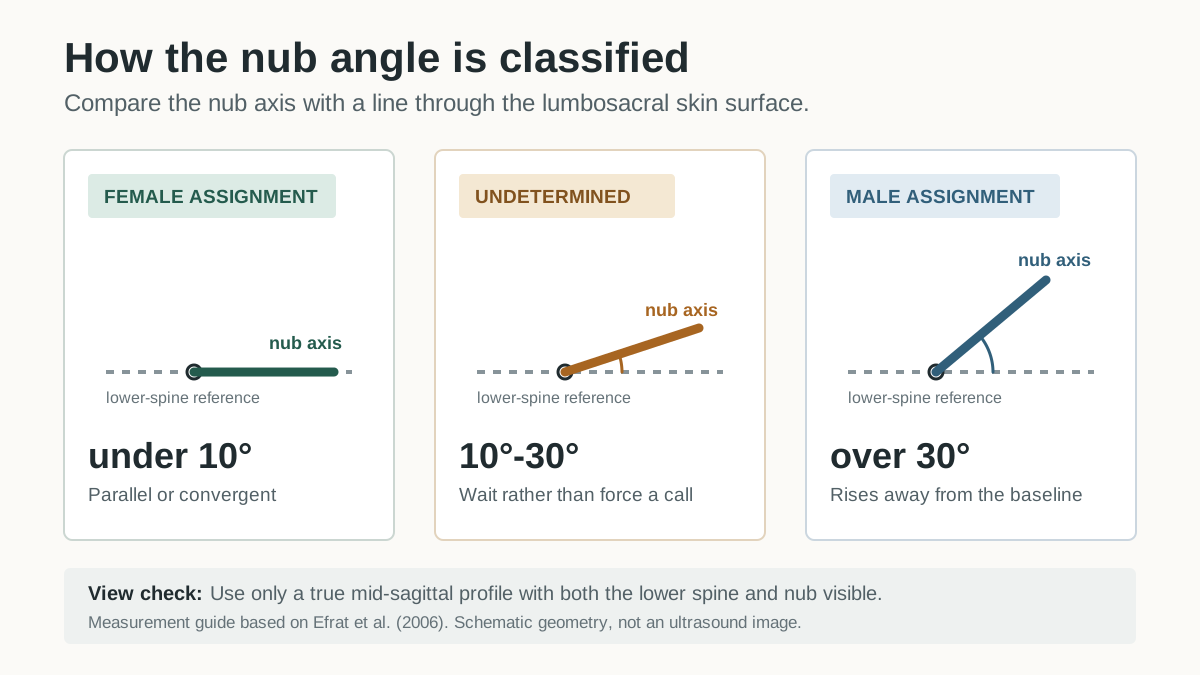
The 10°–30° interval is a real no-call category in the research convention, not a gap to fill with a guess. The hard part is getting a true profile view. An oblique angle can make a boy nub look flat or a girl nub look raised, and that's where most wrong calls come from.
How Accurate Is Nub Theory? The Actual Studies
Nub theory is one of the few pieces of pregnancy folklore with a real evidence base, so here are real numbers instead of vibes:
- Efrat et al., 1999 (the study that formalized the method): sex determination accuracy rose from 70.3% at 11 weeks to 98.7% at 12 weeks and ~100% at 13 weeks, under ideal conditions of expert examiners, good equipment and true mid-sagittal views.
- Efrat et al., 2006 (656 pregnancies, 12–14 weeks): boys identified correctly 99.6% of the time, girls 97.4%. Accuracy for girls climbed with size, from about 91.5% in the earliest 12-week window to ~100% by 13 weeks.
- Hsiao et al., 2008 (11 to 13+6 weeks): overall accuracy around the low 90s, again strongly dependent on gestational age.
Why do we tell parents ~87–92% at 12 weeks when some studies show higher? Those headline figures come from specialists reading ideal images they captured themselves. Real-world material (a phone photo of a printout, a slightly oblique profile, an early-12-week scan) sits below laboratory conditions. Across studies and real submissions, high-80s to low-90s at 12 weeks is the realistic band. Treat anything quoting "99% at 12 weeks" as best-case rather than typical.
Nub Theory Week by Week
Nub theory at 9 weeks
Not readable. The tubercle exists but hasn't begun differentiating, so boy and girl nubs are anatomically identical. A 9-week nub "reading" is a coin flip.
Nub theory at 10 weeks
Still too early. Differentiation is only beginning, and studies don't support meaningful accuracy at 10 weeks. If you can't wait, a blood test (NIPT) already works at this stage. See every method compared.
Nub theory at 11 weeks
The earliest week with any signal, and it's weak: about 70% accuracy in Efrat's data. Better than chance, and wrong almost 1 time in 3, which is poor odds for painting a nursery.
Nub theory at 12 weeks
The classic window, usually coinciding with the NT/dating scan. Expert accuracy reaches ~87–92% (higher in ideal series), provided the image is a clean profile with the nub actually visible. Many 12-week keepsake photos are face shots, which are lovely and nub-less.
Nub theory at 13 weeks
The sweet spot. Multiple series put accuracy at 98–100% by 13 weeks, because the angle difference is bigger and easier to see. The catch is that most parents don't get another scan between 13 weeks and the anatomy scan, so a 13-week nub shot is a lucky bonus rather than something to book.
Boy Nub vs Girl Nub: What the Difference Looks Like
- Boy: the nub rises clearly above the lower-spine reference line. In the cited 2006 study, an angle over 30° was assigned male.
- Girl: the nub lies parallel to or converges with the reference line. In that study, an angle under 10° was assigned female.
The diagram above is intentionally geometric, not a simulated scan. On an actual ultrasound the nub is a few millimeters of gray-on-gray, which is why view quality decides everything.
When Nub Theory Doesn't Work
- Before 11 weeks. No angle has developed yet; there is nothing to read.
- Wrong view. Anything other than a true side profile distorts the angle, and that's the most common source of confident-but-wrong calls.
- Gray-zone angles (10–30°). A real category rather than a failure: some babies haven't rotated far enough yet to call.
- Position and movement. Legs crossed, cord in the way, baby curled. The nub may not be visible at all in your photos.
- Image quality. A photographed printout loses contrast, and the nub is subtle even on a good screen grab.
When the nub isn't readable, the right answer is "unclear," whether it comes from a forum reader, a sonographer or an AI. If someone gives you a confident answer from any photo at all, that's a red flag rather than expertise.
Running Your Own Scan Photo Through the Tool
Our free ultrasound gender predictor applies the method this article describes. On an early-pregnancy profile it looks for the genital tubercle, evaluates its angle against the spine line, then reports boy or girl with a confidence score and the reasons behind the guess. When your image doesn't show a readable nub (wrong view, too early, face-only 3D render) it says "unclear" instead of guessing. Upload your 12-week scan photo and see what it finds. It's for fun rather than a diagnosis, and your photo is never stored.
FAQ
How accurate is nub theory at 12 weeks?
Roughly 87–92% for trained readers on a good profile image at 12 weeks. Some clinical series under ideal conditions report up to 98%, and accuracy climbs toward 100% by week 13. Real-world photos usually sit at the lower end of the range.
Can nub theory be wrong?
Yes. Even at its best it's wrong several times per hundred, and far more often before 12 weeks or on an oblique view. Position, image quality and gestational age are the usual culprits. Only DNA-based tests and the anatomy scan approach certainty.
What is the difference between a boy nub and a girl nub?
A boy's nub angles upward more than 30° relative to the lower spine line; a girl's nub runs parallel to or converges with that line (under 10° in the cited 2006 study).
Can nub theory work at 10 weeks?
No. At 10 weeks the genital tubercle looks the same in boys and girls. Published accuracy only becomes meaningful from 11–12 weeks; before that, a blood test (NIPT) is the only method with real accuracy.
What if the nub angle is between 10 and 30 degrees?
That's the recognized gray zone: the tubercle hasn't clearly committed to either pattern in the image. The right call is to wait, because a later scan, an NIPT result, or the anatomy scan will settle it.
Sources
- Efrat et al. (1999): First-trimester determination of fetal gender by ultrasound
- Efrat et al. (2006): Fetal gender assignment by first-trimester ultrasound
- Hsiao et al. (2008): Fetal gender screening by ultrasound at 11 to 13+6 weeks
- Healthline: Nub Theory — Does the "Angle of the Dangle" Predict Sex?
Got a 12-Week Scan Photo?
See whether it contains a readable nub. Free, instant, and clear when there's nothing to read.
Try the free ultrasound gender predictor
This article is general information, not medical advice. Prenatal testing decisions belong with your OB-GYN or midwife.