3D Ultrasound vs Real Baby: What Can You Compare?
A clear 3D/4D ultrasound can show real facial contours, but it is not a newborn portrait. Only a photograph taken after birth can confirm resemblance. Rather than a factual representation, an AI image is a creative interpretation: it invents surface characteristics missing from the sonogram and might distort or misidentify unclear contours.
This guide covers the information a scan contains, identifies which visible traits are prone to distortion, and contrasts an imaginative depiction with a confirmed post-birth comparison.
Do Babies Look Like Their 3D Ultrasound?
When the baby faces the probe and has fluid in front of the face, a clear scan can depict general facial contours such as the cheeks, chin, lips, and nasal bridge. Although these shapes might seem familiar once the baby is born, sonograms cannot offer a validated similarity rating. The practical timeframe for detailed facial rendering usually spans weeks 26–32 (timing guide).
The scan does not record skin tone, eye color, hair color or photographic texture. Hair may sometimes appear as echoes. While 3D imaging freezes one expression, 4D captures movement across sequential frames. Due to these technical constraints, an ultrasound may share a recognizable silhouette with an infant without looking like a newborn photograph.
3D Ultrasound vs Newborn: What You Can Compare
Analyze the scan feature by feature. A 3D ultrasound renders tissue boundaries as a surface; it does not predict the finished appearance of a newborn face.
| Feature | What may be visible in a clear scan | What the scan cannot confirm |
|---|---|---|
| Nose and lips | Broad profile and contour | Exact size or shape after birth; angle, smoothing and pressure can alter the view |
| Cheeks and chin | Soft-tissue outline | A complete contour when the placenta, uterine wall or shadow blocks part of the face |
| Eyes and eyelids | Eyelid outline and, in some frames, whether the eyes are open or closed | Eye color or how the eyes will look after birth |
| Skin and hair | Surface outline; hair may sometimes appear as echoes | Skin tone, hair color, photographic texture or exact hair density |
This leads to two direct implications:
- Recognizing a similar profile after delivery does not mean the sonogram promised an exact match.
- You cannot judge skin, hair, or family resemblance from the color of the scan, because the orange-gold hue is a visualization setting rather than true skin color.
What 4D Adds to the Same Scan Data
4D adds time to the same volumetric rendering: instead of one still, you watch the baby move. When families later compare the recording with their baby, motion provides more context than a single frame:
- lips moving instead of appearing as one static shape,
- confirmation that a shadow came from the scanning angle rather than anatomy,
- more opportunities to capture an unobstructed, face-forward view.
Motion helps the capture, not the rendering. Each 4D frame uses the same acoustic data as a 3D still, so a session recorded from a poor angle may remain unclear. A short recording still provides more frames to review than one image.
Why the Scan and Newborn Photos Look Different
Four main variables can make comparing a prenatal scan to a newborn picture difficult:
- Newborn swelling. The labor process can temporarily alter the puffiness of the eyelids and cheeks, which is particularly noticeable in snapshots taken right after birth.
- Lighting and lens. Proximity and wide-angle lenses used in delivery room photography can warp facial geometry.
- Expression. While sonograms mostly capture resting faces, newborns cry, squint, and grimace. For a fairer comparison, match a sleeping baby to the ultrasound image.
- Surface detail. Sonograms do not record skin tone, eye color, hair color, or photographic texture. Hair may appear as echoes, but the scan cannot confirm exact hair density. The orange-gold surface is a rendering palette, while realistic details in an AI image are generated.
For a fairer comparison after delivery, choose a calm photo from a similar angle and review several scan frames. A single delivery-room close-up is weak evidence of either a match or a mismatch.
Nose on Ultrasound vs Real: Why It Looks Bigger on the Scan
On a 3D/4D scan, the nose may appear flattened or enlarged because the perspective, physical compression, and software processing alter the depiction:
- Rendering smoothing. Soft-tissue rendering rounds and softens edges, which can make the nose look broader.
- Pressure. Physical contact with the placenta or uterine wall flattens the nasal structure for a short time.
- Angle. Directing the transducer slightly upward overemphasizes the nostrils, mimicking the distortion of a low-angle smartphone picture.
An isolated nose crop cannot separate anatomy from the effects of angle, pressure, and rendering. A clear sonogram is better suited to showing the broad profile; details such as bridge angle, tip shape, and nostril spacing are unreliable when the image is oblique or compressed. Apparent width and puffiness are especially sensitive to position and rendering.
Two additional points about the nose: if its shape varies between frames from the same session, processing and scan angle contribute to the difference, so the most face-forward image is usually the fairest one to evaluate. Baby noses also develop and change after birth, so even a detailed prenatal outline is not a permanent shape.
If the nose is the feature you are focusing on, our dedicated guide to why the nose looks so big on a 3D ultrasound explains the rendering physics feature by feature and what may smooth out after birth.
How Accurate Are 3D Ultrasounds?
Resemblance is not a standard medical measurement, so a 3D ultrasound cannot provide a validated similarity score. Medical sources support a narrower answer:
- The image comes from real anatomy. Prenatal sonography is used clinically to assess anatomy. That medical use does not validate the scan as a tool for predicting facial appearance (ACOG on ultrasound exams, Mayo Clinic).
- Surface appearance is missing. Images are constructed from the reflection of sound waves at tissue interfaces; they do not capture color or photographic surface textures.
- Clarity depends on capture conditions. Gestational week, fetal position, amniotic fluid, equipment, and operator skill all affect the image (which scan type to pick).
View the ultrasound as a structural representation dependent on the scanning angle. Any AI reconstruction derived from it is merely an artistic interpretation, not proof that the baby will exhibit those exact facial traits.
What Affects How Clear Your Scan Looks
Several factors determine whether a facial scan is sharp or obscured: the stage of pregnancy, the orientation of the fetus, the volume of amniotic fluid, the placement of the placenta, alongside the quality of the scanner and the sonographer's proficiency.
Timing is one factor you can plan: the practical window falls within weeks 26–32, which is explained in the week-by-week timing guide. If the face is obscured, ask whether the sonographer can try another position and review the clinic's policy on follow-up sessions.
How Our AI Turns a Scan Into a Baby Photo
The model uses visible scan boundaries as input, then generates a surface the scan cannot capture: skin, hair and open eyes. It may change or misread facial geometry as well as invent surface details, so the result is one creative interpretation, not evidence of preserved features or a prediction of the exact newborn face.
Our 8K ultrasound portrait page shows one example of that transformation and explains which parts come from the sonogram and which details the AI adds.
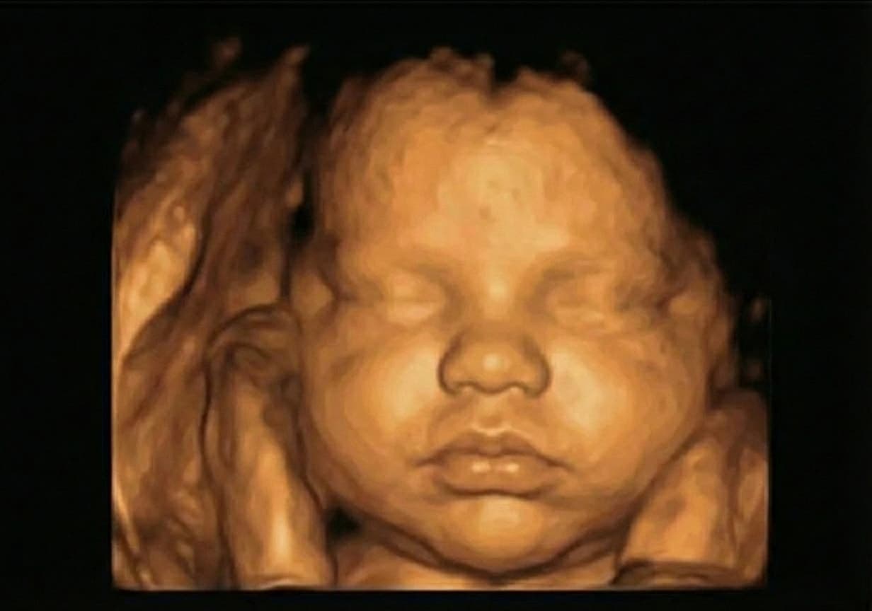 3D scan · 28 weeks
 AI photo from this scan
If you have a scan on your phone right now, you can turn your ultrasound photo into a realistic baby portrait. Portraits are generated after checkout.
FAQ
Do babies look like their 3D ultrasound?
A clear 3D ultrasound can show real facial contours, and families may recognize them after birth. There is no validated resemblance percentage, and scan angle, pressure, and rendering can distort the apparent shape.
Why does my baby's nose look big on the ultrasound?
Rendering angle, pressure against the uterine wall, and soft-tissue smoothing can exaggerate nose width on 3D/4D scans. Tip shape and nostril spacing may be easier to inspect in a clear view, but neither is guaranteed.
Does 4D ultrasound show more facial context than 3D?
4D is not always clearer in a single frame, but real-time motion gives you more chances to capture a clear, face-forward angle.
What week gives the clearest ultrasound face?
Most parents get the clearest face images between weeks 26 and 32. See our week-by-week timing guide for details.
Can AI improve my ultrasound image?
Using an ultrasound scan as a template, AI can produce a realistic-looking rendering. Because it can alter facial proportions and estimate or invent missing details, it is a creative interpretation rather than a medical tool or an actual newborn photograph.
Is this a medical result?
No. It is a creative entertainment experience, not a diagnosis or medical advice. For scan safety questions, see what FDA, ACOG and WHO say about 3D/4D ultrasound.
Trusted Medical Sources and Further Reading
- ACOG: Ultrasound Exams
- Mayo Clinic: Fetal Ultrasound
- WebMD: Pregnancy Ultrasound
- Wikipedia: Obstetric Ultrasonography
For entertainment purposes only. Not medical advice and not a medical diagnosis.